 |
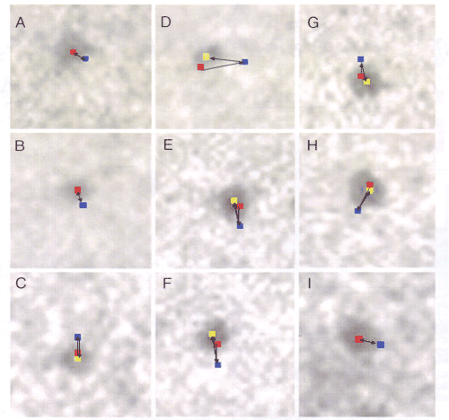
| Figure 11: ecords showing sequential changes in position of 9 different pixels (each 2.5 x 2.5nm) where the center of mass positions of corresponding different particles are located. In each frame, the pixel positions are recorded 3 times, i.e. before ATP application, during ATP application, and after exhaustion of applied ATP. Changes in position of pixels in the first (red), second (blue), and third (yellow) records indicate changes in myosin head position before ATP application, during ATP application, and after exhaustion of applied ATP. Directions of each myosin head movement is indicated by arrow. Note that the myosin heads return toward their initial position after exhaustion of ATP [24]. |