|
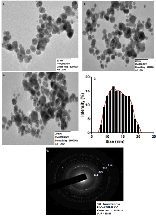
| Figure 3: (A) TEM micrograph recorded from a drop-coated film of gold aqueous solution formed by the reaction of 1 mM HAuCl4 with 1% Padina gymnospora leaf broth at pH 9.5, (B) at 5% leaf broth concentration (C) 10% % leaf broth concentration. The scale bar corresponds to 20 nm, (D) Gold nanoparticles size distribution histogram, (E) Selected area of electron diffraction pattern recorded from one of the gold nanoparticles. The diffraction rings have been indexed with reference to fcc gold. |