|
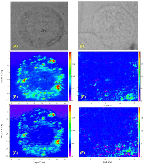
| Figure 2: Spherical GNP as SERS tag: (A) White light image of an A431 cancer cell incubated with 30 nm anti-EGFR labelled GNP; (B) SERS image of the cancer cell obtained by an intensity map of the 1583cm-1 peak; (C) SERS image of the cancer cell obtained by an intensity map of the 1450cm-1 peak; (D) White light image of an NHBE normal cell incubated with 30 nm anti-EGFR labelled GNP; (E) SERS image of the normal cell obtained by an intensity map of the 1583cm-1 peak; (F) SERS image of the normal cell obtained by an intensity map of the 1450cm-1 peak [18]. |