|
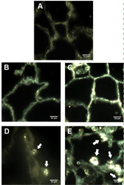
| Figure 3: Darkfield microscopy of lung histology. Representive images of enchanced darkfield microsopy of lung tissue. Naïve male Sprague-Dawley Rat (A), 1 day following citrate vehicle instillation (B), 7 days following instillation of citrate vehicle (C), 1 day following instillation of 20 nm AgNP (D), 7 days following instillation of 20 nm AgNP (E). White arrows indicate punctate formations of AgNP visible in sections. White bar identifies 10 μm scaling. |