|
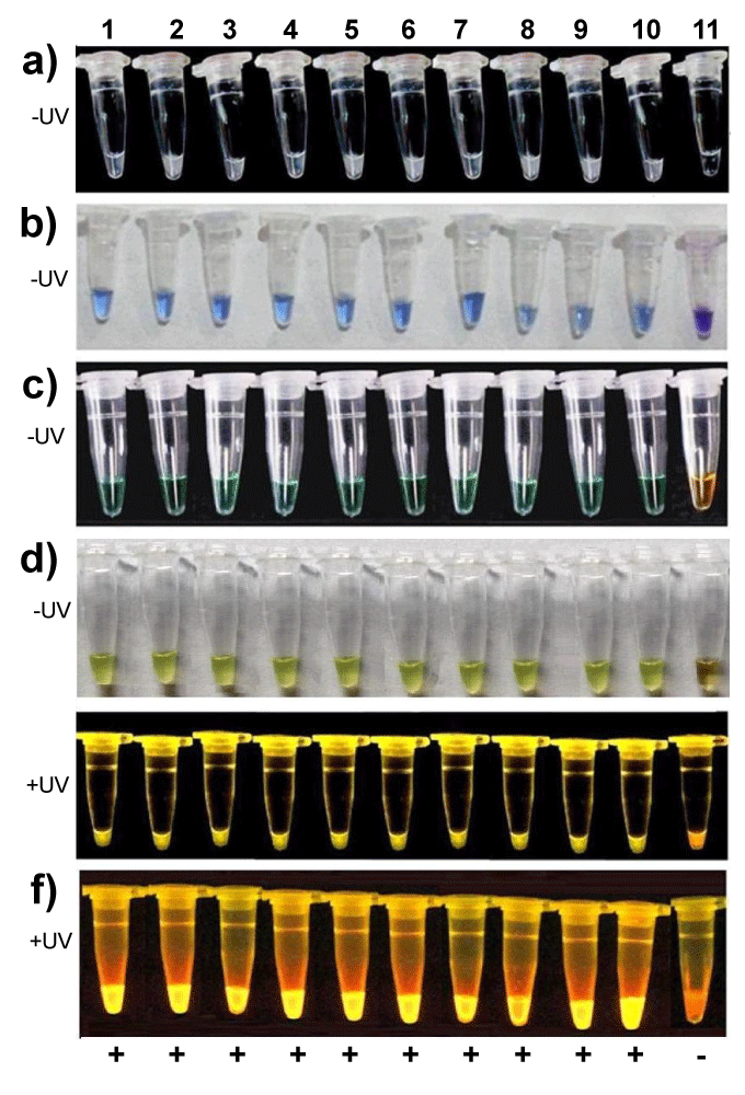
| Figure 4: Detection of positive LAMP reactions using five visualizing methods (different dyes) (a) calcium chloride-based method; (b) hydroxynaphthol blue (HNB)-based method; (c) GeneFinder™ based method; (d) SYBR Green I-based method; (e) SYBR® Premix Ex TaqTM IIbased method; (f) ethidium bromide-based method. Left to right: tubes 1-10, positives samples, respectively; tube 11 negative control (water). |