 |
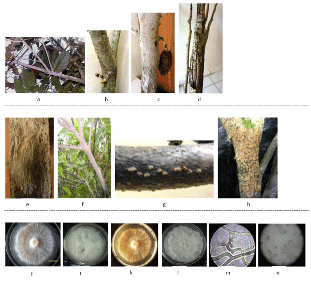
| Figure 2: Symptoms of pink disease (top panel) showing pinkish colouration of infected branches (a), desiccated flowers (b), mummified pod (c) and severe cracks on bark of declining infected cacao tree (d); development stages of E. salmonicolor (Berk. & Broome) on cacao (middle panel) indicating vegetative mycelia mat (e), pink to salmon-colored pustules (f), creamy pustules (g) and orange pustules on a branch (h); and colony morphology and microscopic examination of E. salmonicolor (Berk. & Broome) isolate (lower panel) showing colony morphology 7 dai (i) and 28 dai (j) from cobweb growth stage and 3 dai (k) and 7 dai (l) from pink-coloured pustules, incubated at 28°C ± 2°C on selective medium for basidiomycetes and hyaline non-septate hyphae (m) and irregularly shaped hyaline, unicellular, ellipsoid spores (n) from pustules on naturally infected branches. |