|
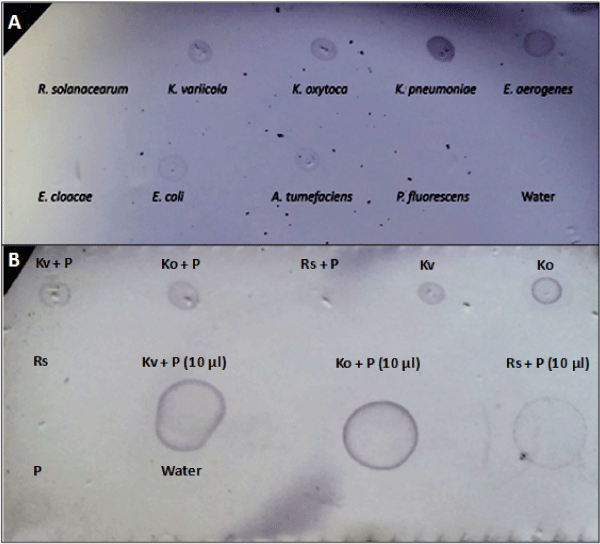
| Figure 2: Colony-blot immunoassay using a Klebsiella-specific antibody. Nitrocellulose membranes were spotted with (A) 1 µl bacterial suspension (ODA600=0.1) or (B) spike plant samples and allowed to dry. A colony-blotimmunoassay was performed on membranes using a Klebsiella-specific antibody. Secondary antibody was a goat anti-mouse antibody conjugated to alkaline phosphatase. Membranes were developed with 1-Step NBT/ BCIP, which yields a purple color as a positive reaction. A faint spot is visible for the 10 μl Ralstonia-spiked plant sample; however that was attributed to background staining of the membrane by plant tissue itself, as seen by the faint spot for the non-spiked plant sample. Kv: Klebsiella variicola; Ko: Klebsiella oxytoca; Rs: Ralstonia solanacearum; P: plant extract. |