 |
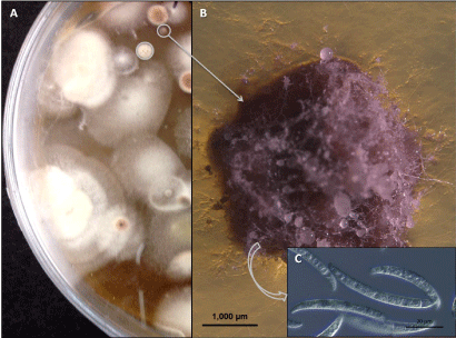
| Figure 2: A representative culture plate of modified Nash and Snyder’s medium (MNSM) showing potential colonies of Fusarium virguliforme (A), photomicrographs (1000 μm) of F. virguliforme colony after subculturing on one-third strength PDA from MNSM culture plate (B), and macroconidia (20 μm) of F. virguliforme (C). Photomicrographs 2B and 2C were taken with an axio V16 florescence stereo zoom microscope and a differential interference contrast microscope, respectively. |