|
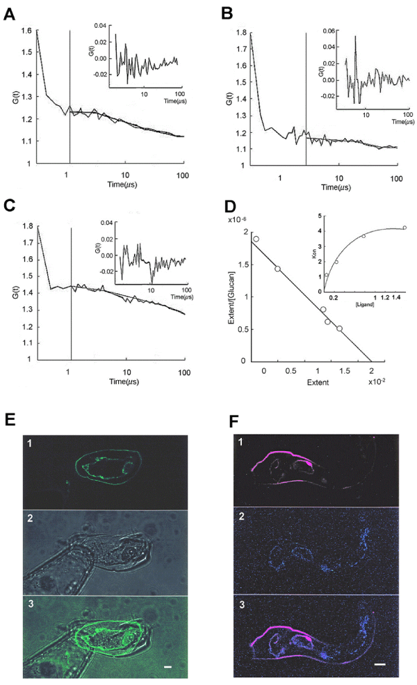
| Figure 3: (a) Fluorescent cross correlation spectroscopy measurements of GFP-CDPK1 and the suppressor glucan with rhodamine-anti- suppressor antibodies (Abs) in potato suspension cells, cv. Rishiri (R1-gene). Typical auto- and cross-correlation spectroscopy of the GFP-CDPK1 and the suppressor glucan. Measurement positions are indicated by the cross-hair (+) in the green channel laser scanning microscopy (LSM) images of GFP-CDPK1 and the rhodamine suppressor in a potato cell. The potato cells were treated with suppressor and then anti-suppressor monoclonal Abs (see Figure 3c). The control treatment without suppressor and the antisuppressor Abs were also tested (data not shown). (b) FCCS analysis showed that potato cells were treated with suppressor and anti-suppressor monoclonal Abs. (c) The potato cell was treated with suppressor and then anti-suppressor Abs with rhodamine anti-mouse monoclonal Abs. (d) Schatterd plots of the binding between suppressor and His-CDPK. Suppressors were at 0,25,50,75, and 100μM. (e) The smGFP-CDPK1 overexpressing potato cells were observed by using green filter of laser scanning microscopy. 1:smGFP-CDPK1 was localized on to the cell wall and plasma membrane of potato cell. 2: Light microscope observation of potato cell. 3: The same images in a potato cell (GFP images were filed up in No.2 photo). (f) The smGFP-CDPK1 overexpressing potato cells were treated with the suppressor of Phytophthora infestans(Pi) and anti-glucan Abs. Green color showed smGFP-CDPK1 proteins. Red colors showed the rhodamine labeled anti-suppressor Abs. |