|
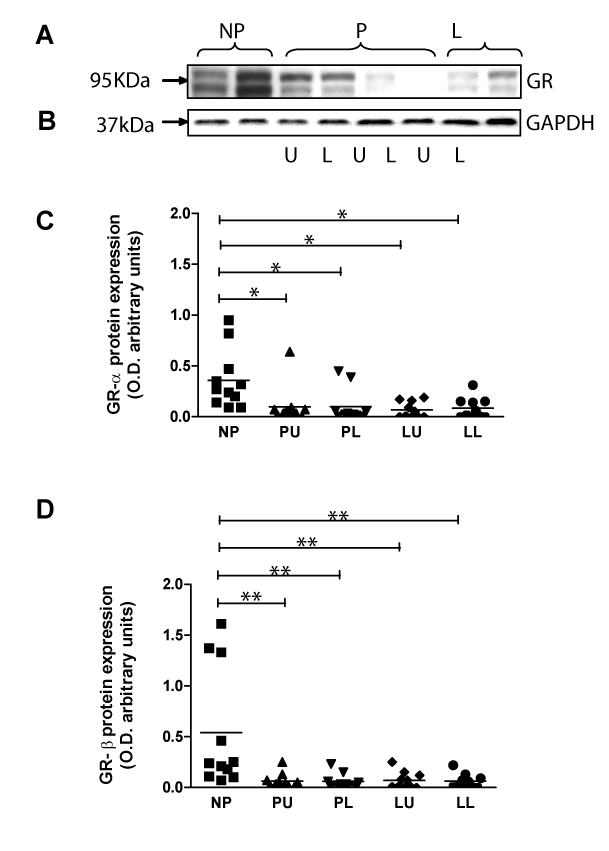
| Figure 2: Expression of Glucocorticoid receptor (GR) protein in human myometrium. Immunodetection of GR protein expression in non-pregnant (NP), uterine upper (U) and lower (L) segments from term non-labouring (P) and term spontaneously labouring (L) human myometrium. Tissue homogenates were resolved by SDS-PAGE and the proteins detected using the BD laboratories anti-GR antibody. Two protein bands at 95 KDa (GR-α) and 90KDa (GR-β) were detected. A representative immunoblot is presented in A. To ensure equal lane loading and transfer efficiency during Western blotting, the membranes were stained with Ponceau-S solution (not shown) and later re-probed with anti-GAPDH as an additional control. A representative control blot is shown in B. Immunodetected bands were quantified by scanning densitometric analysis; densitometry graphs for GR protein are shown in C. All samples are plotted within the allotted patient groups. The bars represent means. (GR-α protein: NP vs. PU/PL/LU/LL P<0.05; GR-β protein: NP vs. PU/PL/LU/LL P<0.01). The results are representative of the samples being used in three independent experiments. D. Western blot analysis of NP tissue homogenate resolved by SDS-PAGE with (+) and without () shrimp alkaline phosphatase (SAP) pre-treatment, with the control shown alongside. |