 |
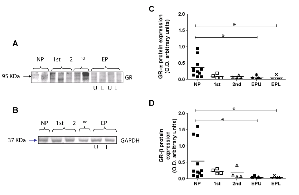
| Figure 3: Pregnancy induced changes in GR expression in human myometrium. Immunodetection of GR protein expression in non-pregnant (NP), first trimester (1st), second trimester (2nd), and preterm labouring (EP) human myometrium. Upper (U) and lower (L) uterine segment samples were examined in the preterm labouring group. Tissue homogenates were resolved by SDS-PAGE and the proteins detected using the BD laboratories anti-GR antibody. Two protein bands at 95 KDa (GR-α) and 90KDa (GR-β) were detected. A representative immunoblot is presented in A. To ensure equal lane loading and transfer efficiency, membranes were stained with Ponceau-S solution (not shown) and later re-probed with anti-GAPDH as an additional control. A representative control blot is shown in B. Immunodetected bands were quantified by scanning densitometric analysis, and densitometry graphs for GR protein shown in B. All samples are plotted within the allotted patient groups. The bars represent means. (GR-α protein: NP vs. EPU/EPL P<0.05; GR-β protein: NP vs. EPU/EPL P<0.05). The results are representative of the samples being used in three independent experiments. |