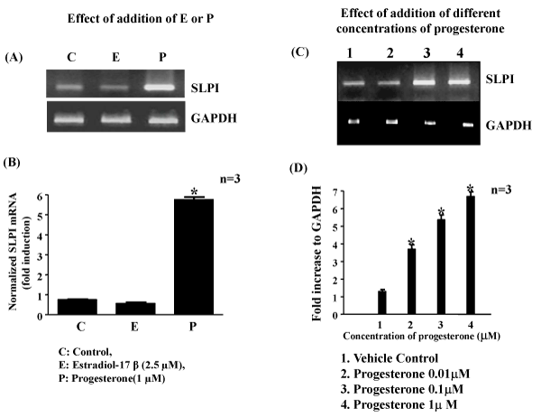
* Significantly different from Control p<0.001.
|
| Figure 1: Regulation of expression of SLPI by steroid hormone in
BeWo cells. A & B: Effect of addition of E2 or Progesterone and C&D:
Different concentrations of progesterone. (A) Cells were incubated
with 1 µM P4 or 2.5µM E2 for a period of 12h. RNA isolated was reverse
transcribed and cDNA was subjected to semi-quantitative PCR in the linear
range of amplification with GAPDH as an internal control. B) Graphical
representation of results presented in A, after normalizing to GAPDH used
as an internal control (values expressed as fold increase over control mean
± S.E. from three independent experiments). C) Cells were incubated with
different concentrations of P4 ranging from 0.01µM to 1µM for 12h. RNA
was reverse transcribed and cDNA subjected to semi-quantitative PCR
in the linear range of amplification. D) Graphical representation of results
presented in C, (values expressed as fold increase over control mean ± S.E.
from three independent experiments).
* Significantly different from Control p<0.001. |