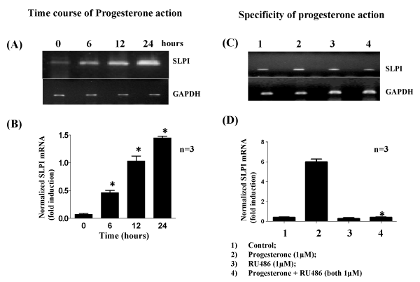
* Significantly different from group 2 p<0.001.
|
| Figure 2: Effect of addition of Progesterone on SLPI expression in
BeWo cells. A & B: Time course and C&D: Specificity of progesterone
action. A) Cells incubated with or without P4 (1 µM) for 024 h. RNA isolated
reverse transcribed cDNA was subjected to semi-quantitative PCR in the
linear range of amplification with GAPDH as an internal control. B) Graphical
representation of results presented in A, (values expressed as fold increase
over control mean ± S.E. from three independent experiments). C) RT-PCR
analysis for expression of SLPI 1) control, (2) P4 (1µM), (3) RU486 (1µM),
(4) P4 + RU486 (both at 1µM) and incubations were carried out for 24 h. RNA
was reverse transcribed and cDNA was subjected to semi-quantitative PCR
in the linear range of amplification with GAPDH as an internal control. D) Graphical representation of results presented in C (values expressed as fold
increase over control; mean ± S.E. from three independent experiments). * Significantly different from group 2 p<0.001. |