|
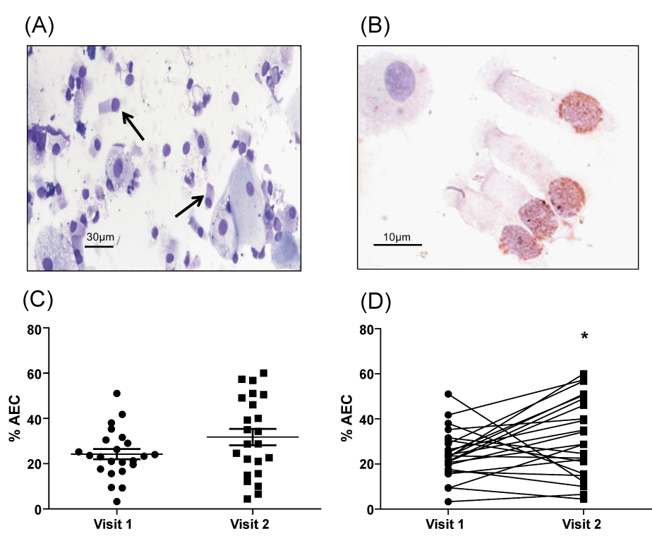
| Figure 2: Airway epithelial cell (AEC) shedding into induced sputum is increased after fluticasone exposure. (A) Representative image of Geimsa stained cytospin demonstrated epithelial cell shedding (arrows indicate bronchial epithelial cells); (B) Representative image of TUNEL staining of shed AEC; (C,D) Difference in the % AEC of total cells counted in the sputum analysis, mean values (C) and paired values (D) at visit 1 and visit 2. (*p<0.05) |