 |
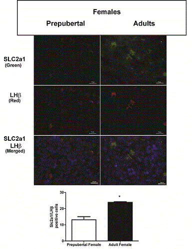
| Figure 3b: Co-localization of SLC2A1 and LH in the murine pituitary gland of pre-pubertal and adult mice. Immunofluorescent analysis of paraffin-embedded pituitary glands isolated from prepubertal and adult C57BL/6J female mice was performed using SLC2A1 and LH antibodies and secondary fluorescent antibodies, with green indicating SLC2A1 positive cells and red indicating LH positive cells. The merge image represents co-localization of SLC2A1 and LH (yellow) with the nuclear DAPI staining (blue). Scale with bar is shown for each image. SLC2a1/LH positive cells were quantified. Graphs representing the average SLC2a1/LH positive cells in pre-pubertal and adult mice of two experiments are shown. Significance was determined by Student’s t-test as shown by * indicating P<0.05. |