|
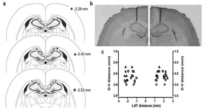
| Figure 1: a Schematic representation of the well placed point of injection illustrating cannulae tip localizations throughout the rostral-caudal extent of the CA1 area of the hippocampus (anteroposterior, -2.3 mm; mediolateral, ± 1.6 mm; dorsoventral, 2.8 mm; from Bregma). Each symbol refers to different anteroposterior distance from Bregma according to Paxinos and Watson Atlas (1998; ○:n=11; Δ:n=19; ◽:n=10). b Representative photomicrographs of half coronal section, cresyl violet-stained, showing unilateral cannulae tip placements (left or right; magnification x2). c Cannulae distributions according to dorsoventral (D-V) and mediolateral (LAT) distance in mm. |