 |
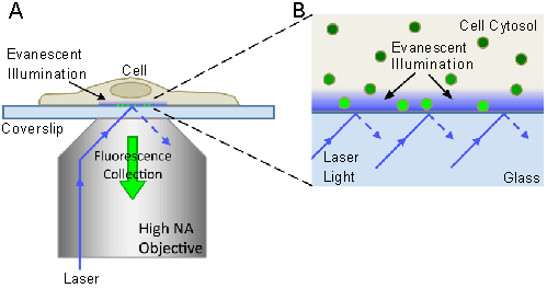
| Figure 1: Through-the-objective TIRFM illumination and detection. (A) Laser light is directed through a high NA objective to the glass coverslip such that it is incident on interface at an angle greater than the critical angle, resulting in total internal reflection. This generates an evanescent wave that illuminates a short depth (~100 nm) into the medium (cell). Fluorescence excited by evanescent illumination is collected through the same objective. (B) Magnification of the cell-coverslip interface region in (A). The evanescent light intensity decreases exponentially with depth. Only fluorophores within the depth of the evanescent illumination field are excited (bright green particles). Fluorescent particles far from the coverslip are not excited and thus do not fluoresce. Figures are not to scale. |