|
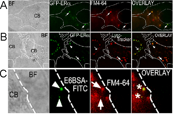
| Figure 3: GFP-ERα colocalize with fluorescent endocytosis markers and E6BSA-FITC in TIRFM. (A,B) GFP-ERα are present on and around the plasma membrane of N-38 neurons. Overlay of FM4-64 (A) or Lysotraker Red (B) fluorescence reveals co-localization of GFP-ERa with vesicles, endosomes, and lysosomes. (C) E6BSA-FITC is present along with FM4-64 fluorescent puncta in N-38 neurons (image C is a close-up taken from a 100× image; B, C are 100× images). BF: Bright field, CB: cell body. |