|
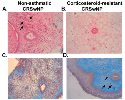
| Figure 1: Nasal polyp sections showing submucosal glands from (A,C) nonasthmatic and (B,D) corticosteroid-resistant CSRwNP patients were stained with haematoxylin (A,B) and Masson’s trichrome stain (C,D). Haematoxylin stain differentiates the type of inflammatory cellular infiltrate in nasal polyps, distinguishing between: (A) major eosinophilic infiltrate (black arrows) in nonasthmatic CRSwNP patients compared to (B) absence of eosinophilic infiltrate in corticosteroid-resistant CRSwNP patients. Masson’s trichrome stain evidences the presence of extracellular matrix and collagen deposition (blue), distinguishing from cellular cytoplasm (pink) and nuclei (dark pink). Images show the differences in fibroblast and extracellular matrix abundance, between (C) non-asthmatic CRSwNP, with scarce presence of fibroblast foci, compared to (D) corticosteroid-resistant CRSwNP, with abundant presence of fibroblast foci and extracellular matrix deposition (black arrows). Representative images are shown. Scale bar=50 μm. |