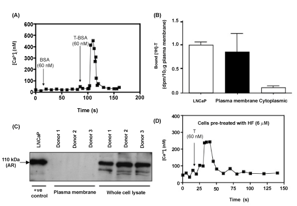
(A) Human endothelial cells (HUVEC) were stimulated with BSA (vehicle control) and then T-BSA (60 nM). Arrows indicate the time of addition of steroid. Buffer was continuously perfused through the chamber during all experiments. Data shown is the average mean curve of 15 cells from 4 separate endothelial cell donors.
(B) Specific binding of [3H]-testosterone to plasma membranes of endothelial cells (HUVEC) or prostate cancer cells (LNCaP). Plasma membranes (10 µg) were incubated at 4°C for 90 mins with 1 nM [3H]-testosterone. Nonspecific binding was determined in the presence of unlabeled T (10 µM), and specific binding represents total minus nonspecific binding. Data is presented relative to LNCaP AR positive control cells and represents the mean of 3 experiments, each performed in triplicate. #P < 0.05 vs. StD; *P < 0.05 vs. HFD.
(C) Western blot analysis for AR protein expression in plasma membrane preparations from human endothelial cells. Plasma membrane (100 µg) and whole cell lysate (20 µg) extracted from HUVEC were analyzed by Western blotting. AR protein was detected in prostate cancer cells (positive control, LNCaP) an whole cell lysates from endothelial cell donors but not in plasma membrane.
(D) Cells were pre-incubated with hydroxyflutamide (HF; 6 µM) for 30 mins and then exposed to T (60 nM). Data shown is the average mean curve of 15 cells from 4 separate endothelial cell donors.