|
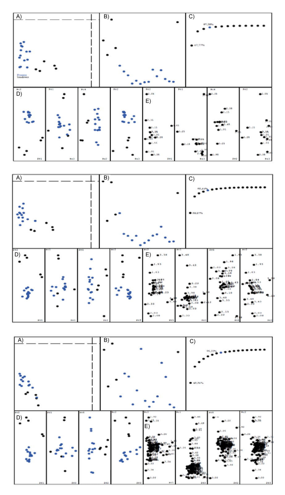
| Figure 2: Figure 2: PCA models obtained using different bucket widths, 0.1 ppm, 0.05 ppm and 0.01 ppm (top to bottom). All samples are color coded using black for healthy and blue for infected. A) Confidence plot for the model, showing that all samples used are well described with the model. B) Deviation plot, showing the distribution of the distances of the samples from the model. For the model with the bucket width 0.01 ppm a equal distribution for healthy and infected is observed. C) Number of PCs explaining the data diversity, values shown indicate variance explained using PC1 and >95% (blue). D) 2D projections plotting the PCs 1×2, 1×3, 4×2 and 2×3. The plots 1×2, 4×2 and 2×3 show good separation of the samples into healthy and infected. E) 2D projections plotting the buckets influence on the localization of samples in the previous plot. Whilst most buckets accumulate on one point, 8 can be identified that are responsible for the separation seen in the previous plot. |