 |
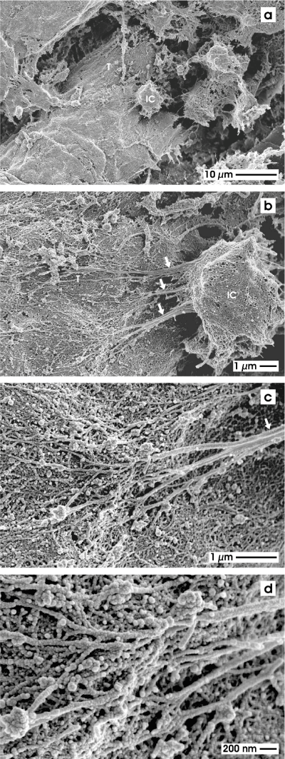
| Figure 7: Scanning electron microscopy of an interstitial cell (IC) contacting a generated tubule (T) by cellular protrusions (thick arrow) within Posi-5 fleece (a, b). Higher magnification shows protrusions which are interwoven with the basal lamina of the tubule (c, d). An exact transition from a cell protrusion to the lamina fibroreticularis at the basal lamina cannot be recognized. |