 |
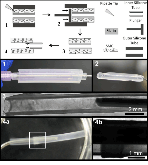
| Figure 1: EML fabrication. Longitudinal view of EML fabrication steps. 1. SMCs are entrapped in fibrin between an outer silicone mold and an inner silicone tube. 2. Fibrin-entrapped SMCs and inner silicone tube were extruded from the mold with a plunger. 3. SMCs are cultured in fibrin for 14 days to form EMLs. 4. EMLs are isolated from the inner silicone tube using forceps. Spontaneous delamination from the inner silicone tube allows for simple isolation of EMLs with forceps (4a, b). Longitudinal retraction occurs concurrent with delamination (4a, b). |