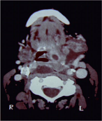
Figure 2:
Axial contrast enhanced-CT at the oropharyngeal level that shows an enhanced tonsilar lesion with affectation of ipsilateral base tongue. Also a 1 cm enlarged lympbh node can be seen.