|
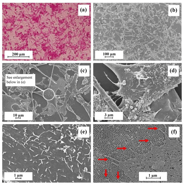
| Figure 3: a) Optical microscopy of MDCK cells on in-house AAO membrane; b) FESEM image of cells on in-house AAO membrane; c) and d) detailed image of cells on AAO membrane; e) enlargement of cell membrane showing microvilli; and f) enlargement of cell membrane showing the presence of filopodia (red arrows). Scale bars shown on each image. |