 |
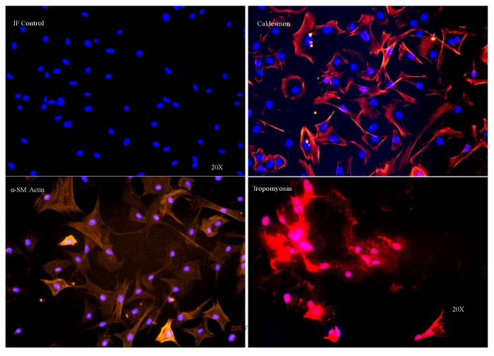
| Figure 1: Phenotypic confirmation of primary vaginal cells. Cells isolated by enzymatic digestion of vaginal biopsies were proliferated in DMEM/12 containing 20% v/v FBS. Cells positively labeled for various SMC markers, such as caldesmon (Alexa Fluor 594, red), a–SM actin (Alexa Fluor 555, orange), and tropomyosin (Alexa Fluor 594, red) confirming the SMC phenotype. Nuclei were labeled with DAPI (blue). |