|
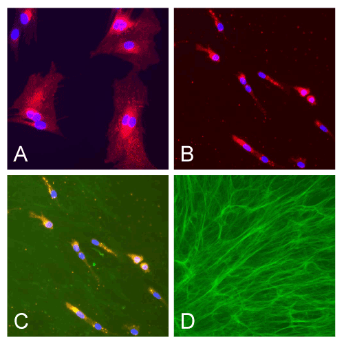
| Figure 2: Dil-stained chondrocytes (red) with DAPI as a nuclear counterstain (blue) were expanded on either Plastic (A) or DSCM deposited by SDSCs (B). DSCM plated with (C) or without the stained porcine chondrocytes (D) were immunostained with collagen I antibody (green). See reference 43 for details. |