|
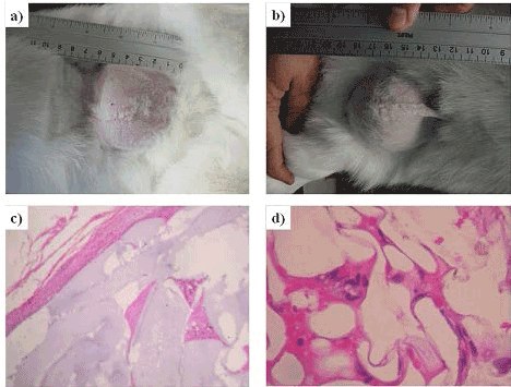
| Figure 7: Images of the surgical area after 2 months (a) and 2 years (b), respectively. Pathology images of the tissue formed around the implanted scaffold after 2 months (c) and 2 years (d) after the surgery. The images showed the obtained results using the hydrogel PHEC30. |