|
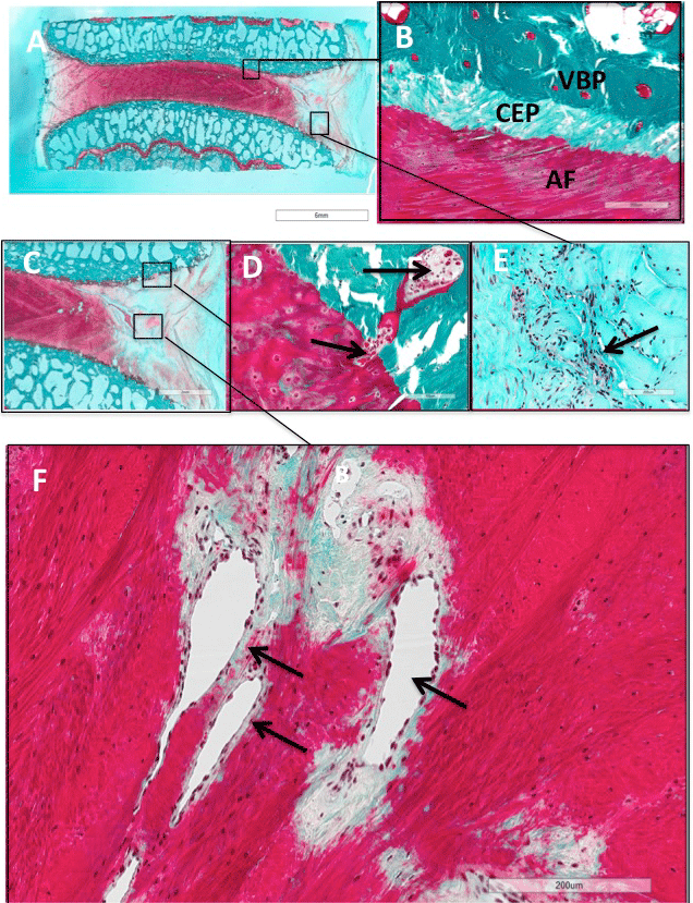
| Figure 8: Examples of histological sections from ovine lumbar discs subjected to surgical incision three months previously. Panel A: Coronal methyl methacrylate embedded section stained with Safranin-O/fast-green showing loss of staining for proteoglycans in the AF region subjected to the 6 x 20 mm horizontal incision. Magnification indicated by 6 mm calibration bar. Panel B: shows a the insertion of the fibres of the AF into the cartilaginous end plate (CEP) and the adjacent vascularized vertebral bone plate (VBP) of an unperturbed region of the disc remote to the site of injury. Magnification indicated by 200 μ m calibration bar. Panels C: Higher magnification of Panel A showing AF lesion regions adjacent to CEP (boxed) and lesion site (boxed. Panel D shows proliferation and invasion of capillary buds of the CEP into the AF (arrowed). Magnification indicated by 100 μ m calibration bar. Panels E and F: shows the presence of capillary vessels at the site of injury (F) and proliferating fibrocytes (arrowed) adjacent to the AF lesion zone. Magnification indicated by 200 μ m calibration bar. |