|
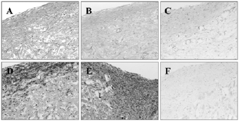
| Figure 4: Microscopic analysis of the tissue engineered leaflets: The histological staining’s (A: HE, B: Elastica van Gieson, C: Masson Goldner, D: alpha smooth muscle actin, E: Vimentin, F: von Figure Legend Kossa) of the tissue engineered heart valve leaflets showed a good cell distribution (A), well developed outer layers but less cellularity in the inner part as well as a good extracellular matrix formation (B). After in vitro conditioning no elastic fibres (C) were detectable. The samples showed a pronounced a-SMA positive layer (D) and vimentin positive cells were detectable (E). Also no calcification was detectable after in vitro cultivation (F). |