|
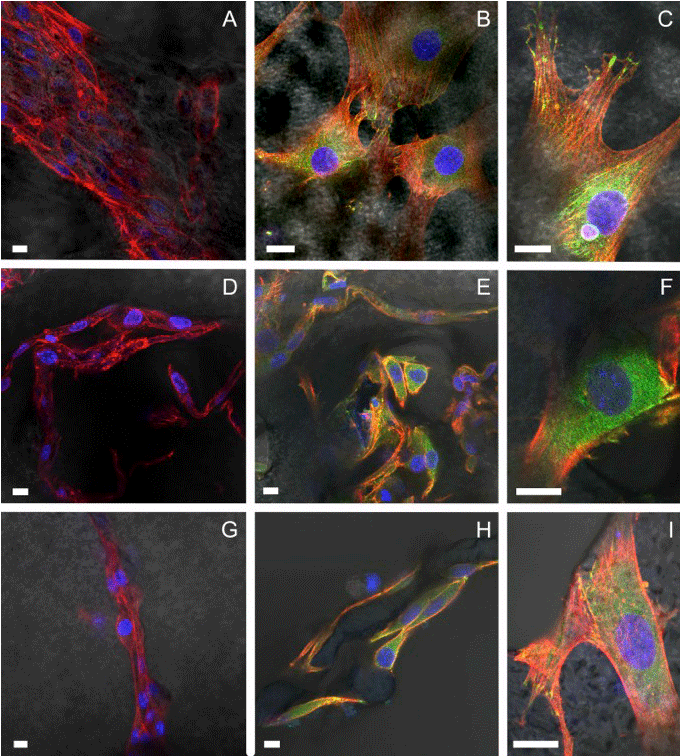
| Figure 4: Confocal fluorescence microscopy of actin (red), vinculin (green) in focal adhesion (yellow in the merged channel) and nuclei (blue) in NIH-3T3 cells. In panels A, B, C was presented the imaging of cells plated on porous scaffold realized without porogens, in D, E, F cells seeded on substrates realized with crystal salts of dimensions <140 μm and in G, H, I images of cells on scaffold realized with porogens of dimension comprised between 140 and 380 μm. Scale bars equals 10 μm. |