|
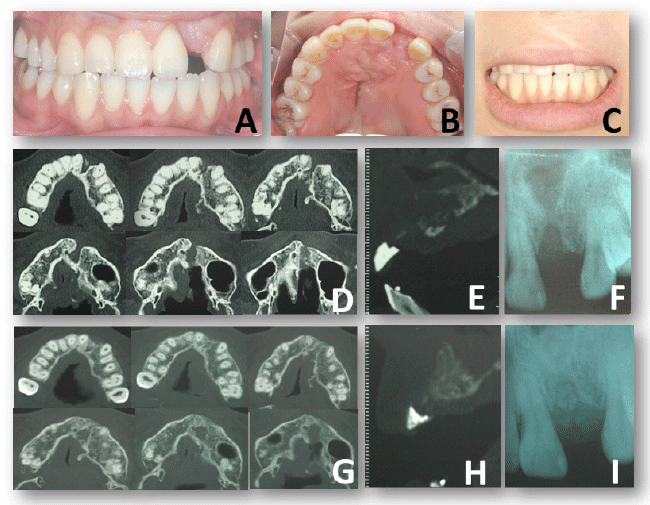
| Figure 7: Description of preoperative diagnosis of a patient with a high-risk factor. The gingival margin of the adjacent tooth showed high scalloped triangle shape compare to the opposite side (A). The width of the edentulous span was appropriate for a tooth (B). The patient has low lip line (C). Computed tomography (CT) before (D, E) and after (G, H) bone grafting for cleft closure. Dental X-ray for evaluation of marginal bone level of adjacent tooth (before bone grafting: F, after bone grafting: I) |