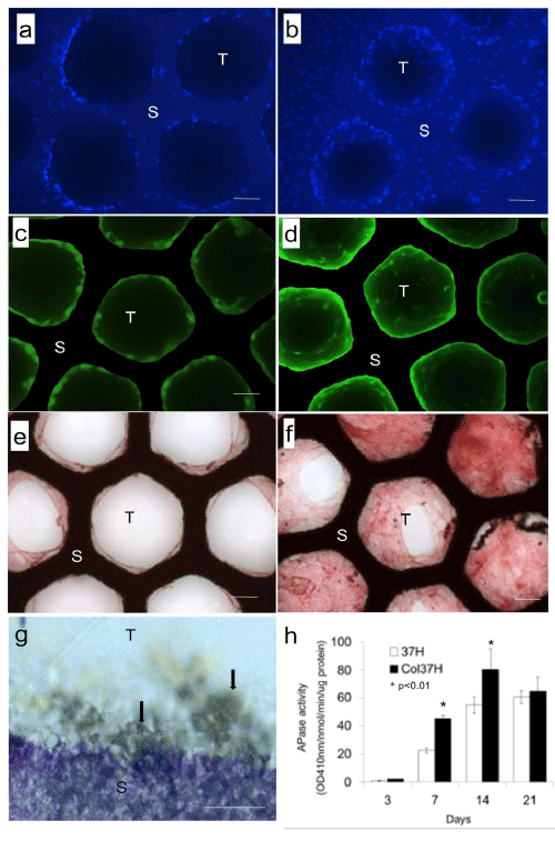
Microscopic findings of MSC cultured on 37H (a, c, e) and Col37H (b, d, f). Samples were stained with DAPI (a and b), type I collagen (c and d), VEGF (e and f) on day 7, and APase (g) on day 14 of the culture. Bars, a-e: 100μm, g: 20μm. The APase-stained cell layer on the inside of the tunnel of Col37H (g, arrows). APase activities were determined on days 3, 7, 14, and 21 of the culture with 37H (◽) or Col 37H (◾) (h). Values represent means ± SE of 6 sets of cultures. Each significant difference was tested between 37H and Col 37H at each time point (*p<0.01) (h). S: Scaffold, T: Tunnel.