 |
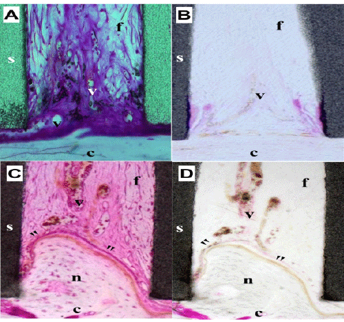
| Figure 8: Microscopic findings on the relationship between blood vessels and new bone formation in a tunnel of Col 37H. Sections were obtained 1 and 3 weeks after implantation. Undecalcified sections were stained with Villanueva bone stain (A and C), and immunological staining was performed with an anti-VEGF antibody (B and D, brown). s: β-TCP, c: calvaria, n: new bone, v: vascular, f: fibrous tissue. Bar: 100μm. |