|
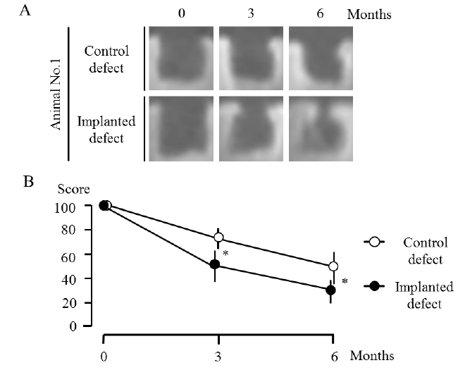
| Figure 2: CT assessment of osteochondral defects. CT images show one cross-section of the multi-planar reconstruction images one, three, and six months after the surgery in animal No. 1 as a representative of all animals (a). Line graph shows the averages of RV (radiolucent volume) percentages at the third and sixth months against those at month zero in both defects (b). The averages at the third and sixth months were significantly (asterisks) decreased in the implanted defects, compared with the control defects. P<0.05 was considered to be statistically significant. |