 |
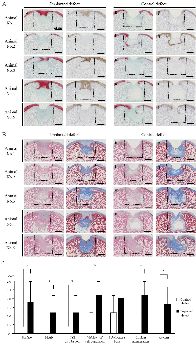
| Figure 5: Histopathology of osteochondral defects using HE, Masson’s trichrome, Safranin O, and immunohistochemical staining of type II collagen (A, B) and microscopic scores of the implanted and control sites with the ICRS histological grading scale (C). In animal Nos. 1, 3, and 4, smooth coverage containing cartilage matrix was positively stained with safranin O and immunohistochemistry (Aa, Ac, Ad, Af, Ah, Ai), and subchondral bone formation was observed on at the implanted sites (Ba, Bc, Bd, Bf, Bh, Bi), whereas the deeply recessed surface and the lower bone formation were left in the implanted sites in animal Nos. 2 and 5 (Ab, Ae, Ag, Aj, Bb, Be, Bg, Bj). At the control site, the surface was irregular and fibrous tissue (which is not stained by safranin O) covered the subchondral bone (Ak, Al, Am, An, Ao, Ap, Aq, Ar, As, At, Bk, Bl, Bm, Bn, Bo, Bp, Bq, Br, Bs, Bt). The black dotted lines indicate the areas of osteochondral defects immediately after the surgery. The ICRS histologic scores at various points except for subchondral bone were higher in the implanted sites than in the control sites in animal Nos. 1, 3, and 4. Animal Nos. 2 and 5 showed that the cartilaginous smooth coverage as well as the subchondral bone did not progress in both defects; therefore the scores at all of the points did not differ between the defects. The scores of the histologic outcome measures such as surface, matrix, cell distribution, viability of cell population, and cartilage mineralization, and the average was significantly (asterisks) higher in the implanted sites than in the control sites (C). |