|
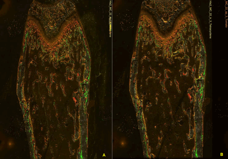
| Figure 5: Image registration using red and green fluorescent beads around the femur. Green and red represent mineralized labels of Calcein and alizarin complexone, and yellow represents TRAP positive cells, orange represents AP stained cells. (A) Overlaid image without registration. TRAP and AP positive cells are not aligned. Note that the file annotation on the right upper corner aligned well. (B) Overlaid image after registration. TRAP and AP positive cells are aligned well with bone matrices. The file annotation on the right upper corner tells how far the TRAP image has been off before registration. |