|
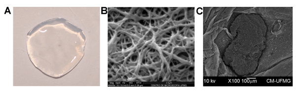
| Figure 4: Morphology analysis of BC membrane and growing iPS cells. Digital photograph of the A) CB membrane surface; B) SEM micrography of the superficial structure of the BC membrane illustrating the disposition of fibers; C) SEM micrography illustrating the morphology of iPS cultured in BC membrane with colony-like structures with defined boundaries. |