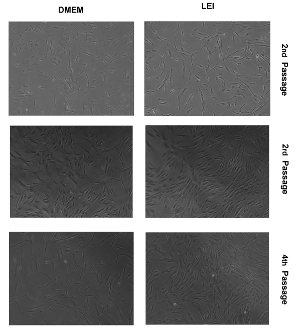
Figure 3:
Images obtained by Optical Microscopy of hASC cultured in DMEM, and adaptation to the LEI from 2
nd
to 4
th
passage.