|
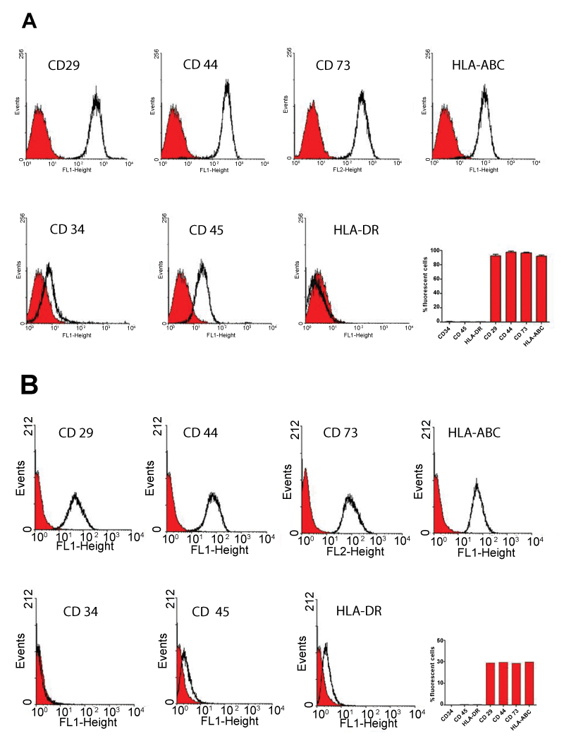
| Figure 4: Phenotypic analysis by flow cytometry of hASC cultured in medium (A) LEI and (B) DMEM. The histograms show the number of events versus fluorescence intensity. The red curve is the fluorescence of the negative control, the black line shows the cell populations evaluated for a specific marker, and its shift to the right indicates the existence of marking. |