 |
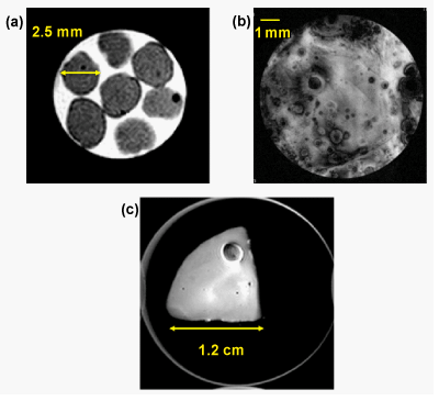
| Figure 2: Examples of MRI images of cartilage and bone engineered tissue constructs. T2 weighted axial slice of: (a) of alginate beads seeded with chondrocytes at 14.1 T. Seven beads are shown in the images indicating that statistically significant results can be obtained in one experiment; (b) Osteochondral tissue constructs (thanks to Prof. Wan-Ju Li at the University of Wisconsin-Madison) with very high resolution at 11.7 T showing dark spots presumably because of bone mineralization; (c) Cartilage monolayer culture purchased from “articular engineering (http://articular.com/)” at 14.1 T. |