|
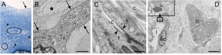
| Figure 6: Characteristics of fibrous RT of transplanted cartilage defects shown by toluidine blue stained semithin sections (A) and TEM images (B,C,D). A-D: Fibrous tissue of the middle of the cartilage defect treated with chondrocytes on a double sandwich-like layer of a hyaluronan web and a collagen type I/III fleece. A: The tissue contains vascular-like structure in the lower part of the RT (circles), many cells (dark blue dots; arrowheads) in deep regions and fewer cells in the upper half. The surface is smooth and locally covered by a dark layer (arrow). SB=Subchondral Bone. B: Part of the cell body and cell extensions of a polygonal chondrocyte embedded in fibrous matrix. Note the organisation of collagen in form of strands (asterisk). A very thin rim of electron-dense matrix (arrow) is locally visible along the cell membrane. The cytoplasm of the cell is partially dominated by dilated rER. C: Detail image of a cell extension of a polygonal cell containing loose strands of Filaments (f) in the cytoplasm and many Vesicles (v) aligned at and budding from the cell membrane. The large, bright fibre in the middle of the cell extension is a Collagen fibril (c) penetrating the cell in a deep invagination (note that it has the same appearance as the collagen fibrils in the matrix surrounding the cell). The fibril is flanked by the cell membrane (arrowheads). D: Vascular-like structure formed by Endothelial Cells (ec; l: Lumen) with dark Weibel-Palade-like bodies in the cytoplasm (see insert). In the matrix surrounding the capillary, a very large mast cell-like cell with densely filled, homogeneous inclusions is visible (ic). Scale bars: (A) 25 µm, (B) 2 µm, (C) 500 nm, (D) 3 µm. |