|
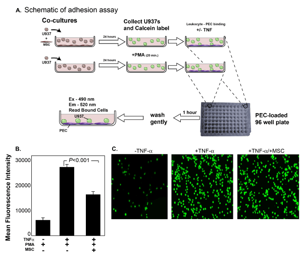
| Figure 1: MSCs decrease functional leukocyte-endothelial cell binding. A. Schematic of Co-cultures: U937 leukocytes were cultured with or without MSCs for 24 hours. The cells were subsequently labeled with Calcein-AM. U937s were stimulated with PMA (200ng/mL) for 20 minutes. Pulmonary endothelial cells (PECs) were stimulated with TNF-α (10 ng/ml)-, 4 hours prior to addition of U937s. 25,000 U937s/well were added to a 96 well plate that was pre-loaded with confluent (PECs) The cells were incubated for 60 minutes and gently washed 3 times with PBS. Adherent cells were quantified by measured fluorescence on a BioTek Synergy 2 plate reader (Biotek, Winooski, VT). B. Binding Assay: TNF stimulation of PECs increased U937 binding (Group 1 vs. Group 2) Binding was significantly decreased in Group 2 which was co-cultured with U937s compared to Group 3. Binding was quantified by mean fluorescence intensity (MFI) on a flourimeter. C. Representative Images of Binding: Qualitative images of representative wells showing decreased binding of fluorescent U937s co-cultured with MSCs to PECs. Images were taken at 20X magnification. |