|
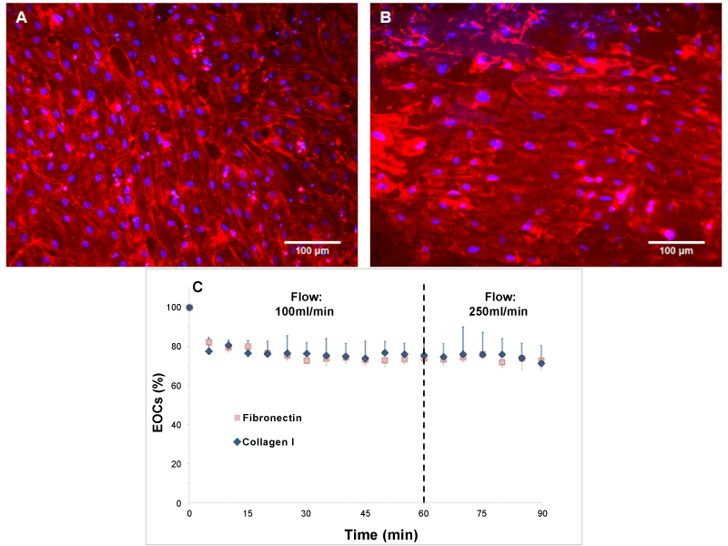
| Figure 4: EOCs on collagen I (A) and fibronectin (B) coated ePTFE graft surfaces after 48 hours. Nuclei stained with DAPI are shown in blue, and the cytoskeleton (f-actin) is stained with Alexa 568 phalloidin shown in red. EOCs attached and spread on the ePTFE modified surface. (C) Durability test of 111Indium labeled EOCs on protein-coated ePTFE in the baboon ex vivo shunt system. There was an initial drop (from static to first 5 min frame) in cell numbers with the initiation of flow, but cells remained stable on the graft through 60 min at standard flow rates used in the ex vivo shunt studies. Data are shown as a percentage of EOCs at time zero for the respective surfaces. |