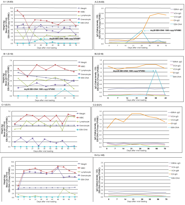
The body weight, number of WBCs, lymphocytes, granulocytes, and EBV-DNA copy (A-1, B-1, C-1, and D-1) are indicated with the time course after EBV inoculation. The EBV-related antibody levels and EBV-DNA copy numbers (A-2, B-2, C-2, and D-2) are shown at the indicated times after EBV inoculation. When the EBV-DNA copy number was lower than the minimum test sensitivity (20 copies/106 WBCs), it was recorded as “0.” Rabbits N-83 (A-1 and A-2) and O-19 (B-1 and B-2) are EBV-DNA-positive rabbits. The data for rabbits O-21 (C-1 and C-2) and L-143 (D-1 and D-2) indicate a PB EBV-DNA-negative rabbit with no detectable EBER-1 expression in tissues and a PB EBV-DNA-negative rabbit with a few EBER-1-expressing lymphocytes detected at necropsy, respectively. For rabbits N-83 and O-19, EBV-DNA was detected transiently, with maximum 1.3×10³copies/106 WBCs on day 28 and 1.3×10³ copies/106 WBCs on day 35, respectively. Rabbit O-19 died on day 42 after a transient increase in PB EBV-DNA copies and a gradual, continuous loss of body weight. The numbers of WBCs, lymphocytes, and granulocytes exhibited no apparent trends following EBV inoculation. Regarding EBV-related antibody levels, none of these antibody levels were increased in the vaccinated rabbits (B-2, C-2, and D-2).In contrast, in the control rabbit N-83high levels of EA-IgG were maintained during the observation period after a rapid EA-IgG increase during the early phase, although no increases in VCA-IgG and EBNA-IgG levels were observed (A-2).