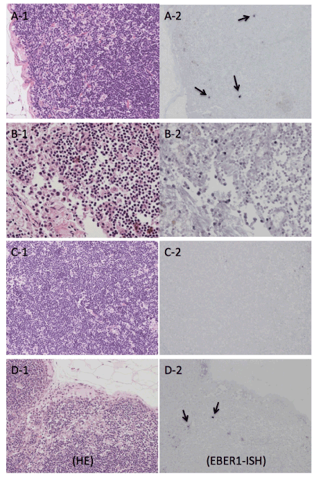
Hematoxylin-eosin stained histologies (A-1, B-1, C-1 and D-1) and EBER-1 expressions (A-2, B-2, C-2 and D-2) in lymph nodes were shown. A-1, C-1, and D-1: No abnormalities were found in HE stained specimens. ISH revealed that a few EBER-1-positive lymphocytes had infiltrated into the lymph nodes of the control and L-143rabbit (A-2 and D-2, arrows), but no EBER-1 expression was detected in the lymph nodes of rabbit O-21 (C-2). For the lymph nodes from rabbit O-19 that died on day 42, there was a trend for degeneration combined with an increase in macrophages (B-1) and an unexpected increase in the number of EBER-1-positive lymphocytes(B-2). Original magnification was×200 in B-1 and B-2, and ×100 in the others.