|
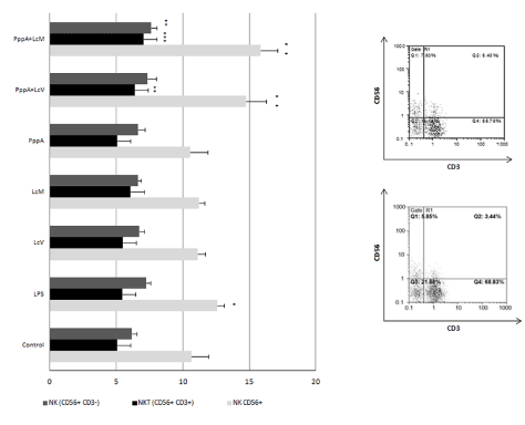
| Figure 1: Effect of live (LcV), heat-killed Lactobacillus casei (LcM), pneumococcal antigen (PppA), combinations of both Lc and PppA (PppA+LcV, PppA+LcM) and LPS on expression of surface markers CD56 and CD3 in peripheral blood mononuclear cells. A) Control and B) PppA+LcM are representatives dot-plot using anti-CD3 and anti-CD56 mAb, allowing identification of 3 cell subsets: CD3-CD56+ (NK cells), CD3+CD56+ (NK-T cells) and CD3+CD56- (T cells). Graphic indicates the percentage of cells for each of these subsets. Data are expressed as mean ± ds values of 4 healthy donors. Duplicate assays were performed for each donor and each stimulus. (*P<0.05, **P<0.01, ***P<0.001). |