|
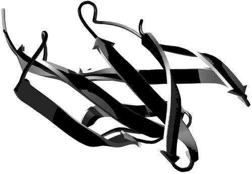
| Figure 7: Structural superposition of the model and target structures. Ribbon representation as executed using the Swiss-PDB Viewer and rendered with the POV-ray. The mold structure of the prM protein of the dengue-2 virus is presented in black (C Chain - code PDB: 3C5X) and the structure model is presented in gray (prM/DENV-3). The RMSD between both carbons is 0.21Å. |