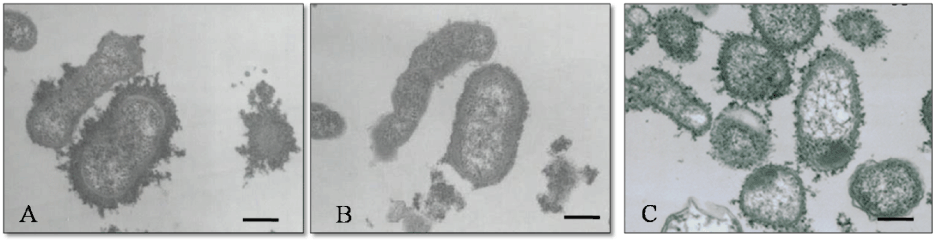
Figure 5:
Electron micrographs of
P. multocida
strains P-1059 (A), P-1059C (B) and Δ
hexB
(C). Bars, 200 nm.