|
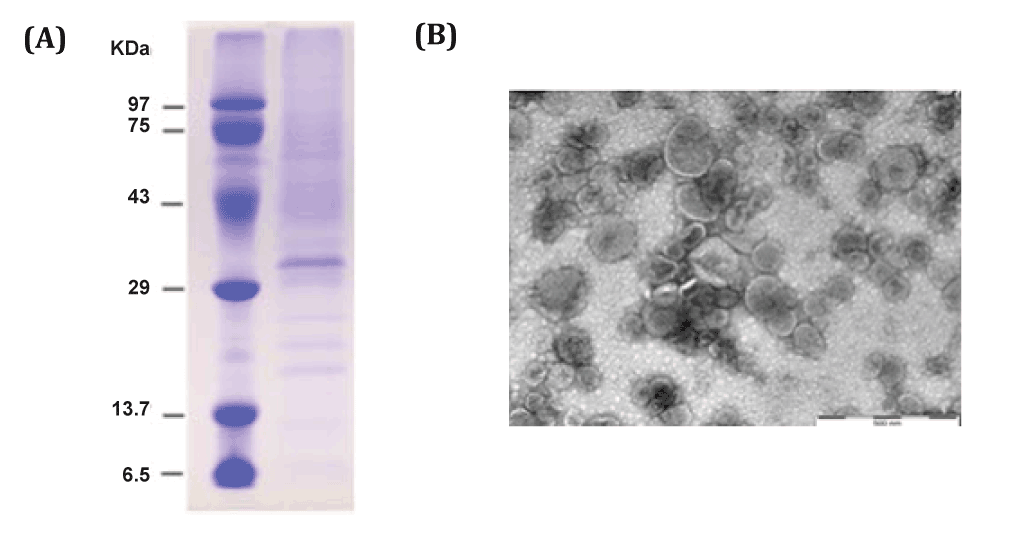
| Figure 1: Characterization of L. interrogans OMVs. (A) 10 μg of purified Leptospira OMVs were loaded into 12% SDS-PAGE. Large numbers of protein bands of variable size were detected. The most distinct band located between 43 and 29 kDa protein markers likely represents LipL32, which is the most abundant outer membrane lipoprotein. (B) A TEM image of Leptospira OMVs shows closed spherical structures 40 to 300 nm in diameter. Bar indicates 500 nm. |